Written by Amanda R. McDaniel, MS, BSN, RN
Amanda is a BSN/RN with a MS in Physiology and a BA in English. She worked as a medical writer in the pharmaceutical industry for 11 years before pursuing a career in nursing. She now works as a nurse on a NeuroTelemetry unit and continues to write and edit on a freelance basis. Amanda’s LinkedIn
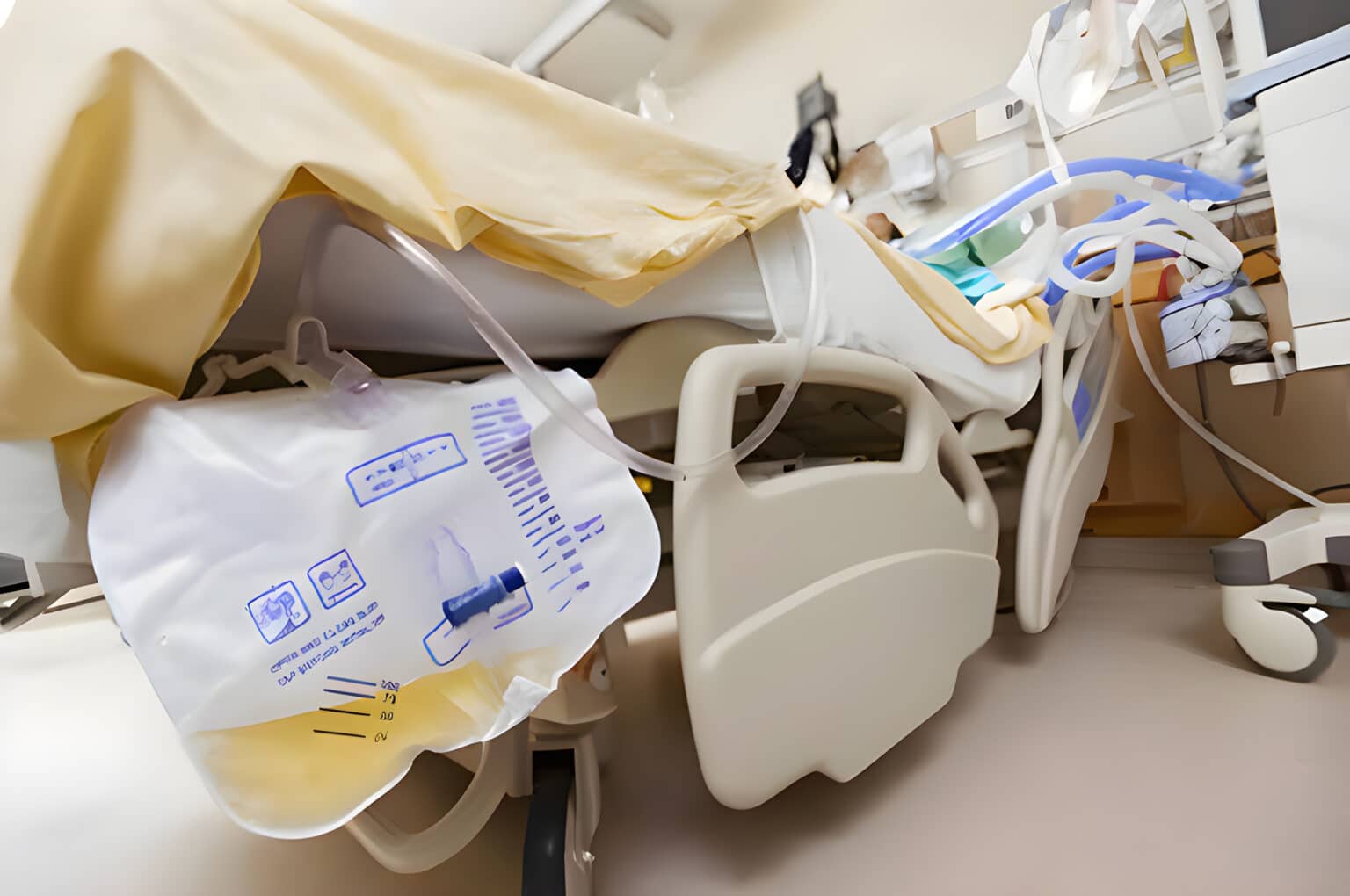
Accurate measurement of urination (aka, the output portion of intake and output) allows medical personnel to assess kidney and bladder function. Changes in output quantity or quality can reflect health status changes including new-onset infection or renal injury.
- Gather your supplies:
- Gloves
- Graduated measuring container. Make sure that the measurement on the container reflects the accuracy required by the doctor or institutional policy.
- Antiseptic wipes
- Paper towels or an absorbent pad
- Give the resident privacy by closing the door or curtain.
- Perform hand hygiene and don gloves.
- Lay the paper towels or absorbent pad on the floor below the urinary drainage bag.
- Place the measuring container on the towels or pad.
- Without allowing the drain to touch any part of the measuring container, open the drain and allow all urine to drain into the container.
- Clamp the drain and clean the end with an antiseptic wipe. Place the drain back in its holder.
- Note the amount of urine in the container. Note the characteristics of the urine. What is the color? Is there sediment or blood present? Does it smell strongly? Is there a decrease or increase in the amount of urine versus the last time the bag was emptied?
- Remove the paper towel or absorbent pad.
- Pour the urine into the toilet and rinse the measuring container. Pour the rinse water into the toilet and flush.
- Disinfect and store or dispose of the measuring container.
- Remove gloves and perform hand hygiene.
- Record the quantity and characteristics of the urine in the appropriate section of the resident’s chart per institutional or unit policy. Report any changes to the nurse per policy.
References
S. A. Sorrentino, & L. N. Remmert. (2012). Urinary elimination. In Mosby’s textbook for nursing assistants (8th ed., pp 399). St. Louis, MO: Elsevier Mosby.